When achieving a perfect smile, orthodontic treatment is often the key. But have you ever wondered how your orthodontist knows if your teeth are moving in the right direction? The answer lies in the power of X-rays. These diagnostic tools play a crucial role in monitoring the progress of your orthodontic journey, ensuring that you receive the most effective and efficient treatment possible.
The Role of X-Rays in Initial Assessment
Before beginning any orthodontic treatment, your orthodontist will thoroughly examine your teeth and jaws. This initial assessment includes taking X-rays, which provide a detailed look at the underlying bone structure, tooth roots, and jaw placement. By analyzing these images, your orthodontist can identify issues such as misaligned teeth, overcrowding, or bite problems that may require correction.
X-rays are particularly useful in detecting problems that may not be visible during a physical examination. For example, they can reveal impacted teeth, which are teeth that have not erupted through the gums and may require special treatment to guide them into the proper position. X-rays can also show the presence of extra teeth or missing teeth, which can impact the overall treatment plan.

Types of Orthodontic X-Rays
Orthodontists use several types of X-rays to gain a comprehensive understanding of your oral health:
- Panoramic X-rays: These X-rays provide a complete view of your mouth in a single image. They show the positioning of your teeth, jaw bones, and any potential issues with your sinuses or temporomandibular joints (TMJ).
- Cephalometric projections: These X-rays show a side view of your head, allowing your orthodontist to examine the relationship between your teeth and jaw and your facial profile. This information is essential for developing a personalized treatment plan.
- Cone Beam CT: This advanced imaging technique offers a three-dimensional view of your teeth, bones, and soft tissues. It provides the most detailed information about your oral structures, helping your orthodontist make precise treatment decisions.
Cone Beam CT is particularly useful for complex cases involving facial asymmetry or severe jaw discrepancies. The 3D images allow orthodontists to visualize the relationship between the teeth, jaws, and surrounding structures from every angle, creating a more accurate and effective treatment plan.

Monitoring Treatment Progress
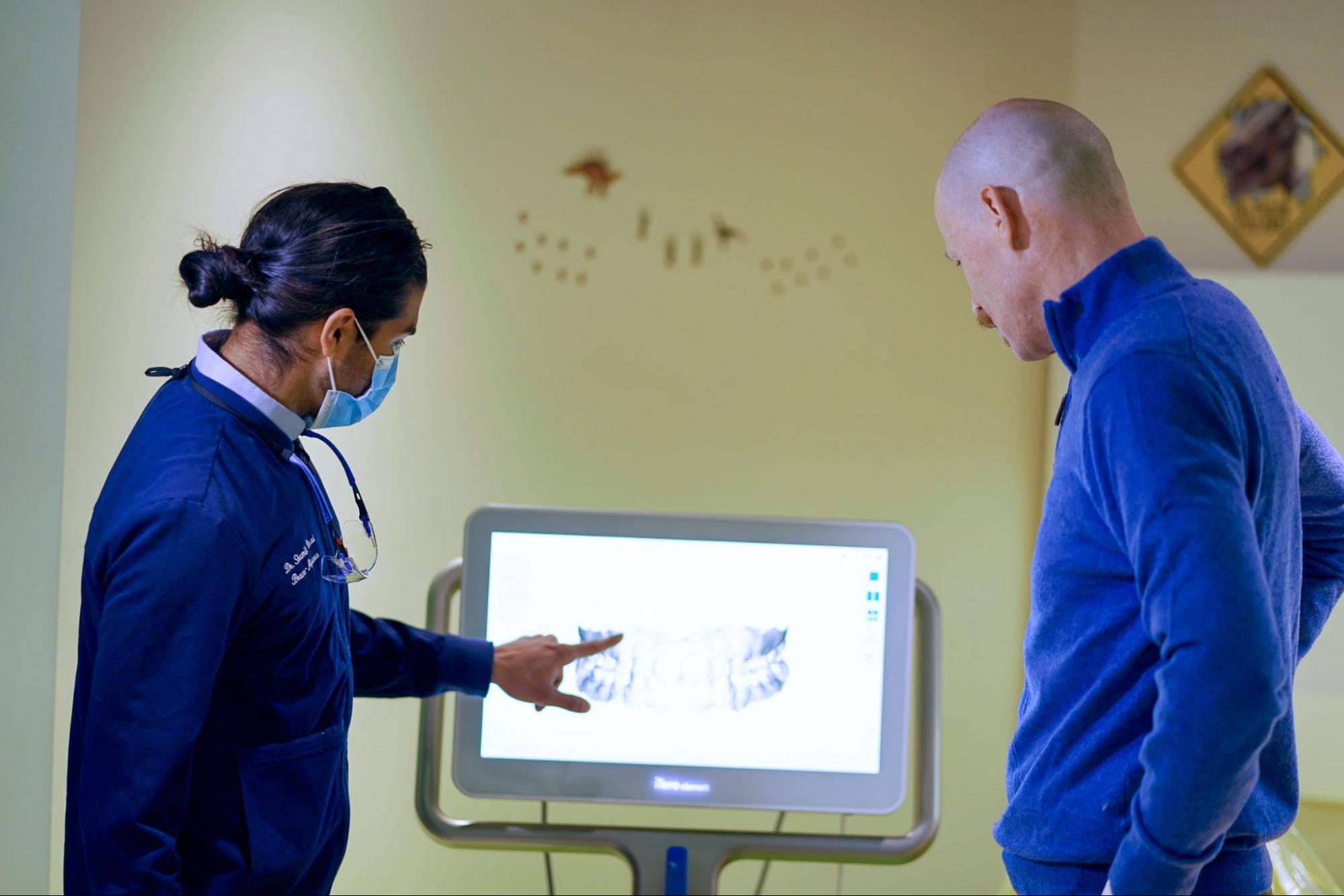
Throughout your orthodontic treatment, periodic X-rays are important to track the movement of your teeth and evaluate the response of your bones to the treatment. These images help Dr. Desai determine if your teeth are moving as planned and if any adjustments to your treatment plan are necessary. By closely tracking your progress, your orthodontist can ensure that you achieve the best possible results most efficiently.
Progress X-rays are typically taken every 6 to 12 months, depending on the complexity of your case and the type of treatment you are receiving. For example, patients with traditional metal braces may require more frequent X-rays to monitor the movement of their teeth, while those with clear aligners like Invisalign may need fewer X-rays due to the predictable nature of the treatment.
Technological Advancements in X-ray Imaging
In recent years, there have been significant advancements in X-ray technology that have greatly benefited the field of orthodontics. Digital X-rays, for example, offer higher-quality images with lower radiation exposure than traditional film X-rays. Specialized software used in orthodontics can now predict tooth movements and treatment outcomes using initial X-rays, allowing for even more precise treatment planning.
One such advancement is using computer-aided design and computer-aided manufacturing (CAD/CAM) technology in orthodontics. This technology allows orthodontists to create digital models of a patient’s teeth and jaws using X-ray images and intraoral scans. These models can then be used to design custom orthodontic appliances, such as clear aligners or custom brackets, tailored to the patient’s needs. CAD/CAM technology improves the accuracy and efficiency of orthodontic treatment and enables orthodontists to provide more predictable outcomes for their patients.
Safety Measures for X-Rays
While X-rays are an essential tool in orthodontics, it’s understandable that there are concerns about radiation exposure. Rest assured that dental X-rays are safe, and orthodontists take numerous precautions to minimize exposure. This includes using lead aprons to protect your body and utilizing modern technology designed to emit the lowest possible amount of radiation while still providing high-quality images.
Orthodontists follow the ALARA principle, “As Low As Reasonably Achievable.” This means they strive to use the lowest radiation dose possible while obtaining the necessary diagnostic information. Furthermore, the American Dental Association (ADA) and the American Association of Orthodontists (AAO) have established guidelines for the safe use of X-rays in dental and orthodontic practices, ensuring that patients receive the maximum benefit with minimal risk.

Achieve the Smile You’ve Been Longing For
At Elm Tree Orthodontics, we understand the importance of X-rays in achieving the smile of your dreams. Our team of experienced professionals utilizes state-of-the-art technology to develop personalized treatment plans that deliver outstanding results. Whether you have a question about how we use X-rays or if you are ready to schedule a consultation, we are excited to help.